Athletic Injuries
Injuries of the knee in athletes vary somewhat
from injuries in other patients. This is due to
several factors. The degree of conditioning and
quality of the tissues in athletic knees are
higher. The types of injury are more sports
specific (for example, anterior cruciate
ligament tears). Finally, the expectations of
the athletes for post treatment functional
outcome tend to be higher due to their sporting
interests. Thus, surgical techniques have
evolved in the last 10 to 20 years that have
allowed for less invasive repair and
reconstruction of knee injuries in the athlete
to allow a high level of return to function with
minimal invasiveness.
Ligament Sprains:

The term "sprain" refers to a partial or complete tear of ligament substance. Ligaments are made up of millions of fibers of "collagen", somewhat like a woven rope. When a ligament is sprained, a portion of the fibers is dredged or torn. In low-grade sprains, only a few fibers are torn, and patients have pain without much in the way of instability from the ligament being too loose. In high-grade or complete sprains, a large portion of the fibers is torn and the ligament ends may be fully separated from either other. In this setting, there will be pain as well as instability of the knee. Low-grade sprains are usually treated with activity modification, physical therapy, anti-inflammatory medications, and time. High-grade sprains (or tears) of ligaments may require a surgical treatment, such as an anterior cruciate ligament tear.
Muscle Strain:

Muscle strains are similar to ligament sprains, except that they occur in muscle tissue rather than in the collagenous ligament substance. This is what occurs, for example, when an athlete has a "hamstring pull". The same rating of muscle strains is used from low-grade strains in which a few of the muscle fibers are torn, and the athlete has pain without much weakness, to high-grade complete tears of the muscle when severe pain and weakness are present. As in ligaments sprains, the treatment is based upon the type of strain and the degree of injury.
Cartilage Tears:

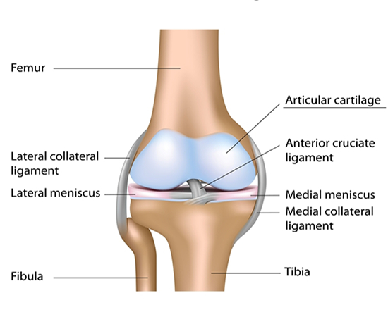
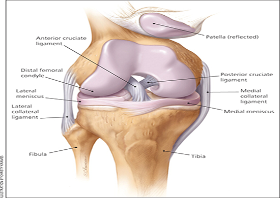
There are two forms of cartilage in the knee.
One is a surface, or "articular" cartilage,
which is the white, glistening cartilage on
the surface of the bone. This is very smooth
and designed to glide
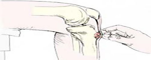
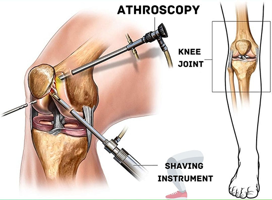
without any
friction. The second type of cartilage is
the "meniscus" cartilage. This is more of a
"bumper cushion" cartilage, which sits
between the thigh and shin bones, providing
a shock-absorbing effect during athletics.
When the knee is violently twisted or other
injuries occur, the meniscus cartilage can
be torn. If this is a small tear and does
not have much in the way of symptoms, it can
be treated without surgery. If it is a large
tear, with a loose fragment that flips up
into the joint and blocks or limits motion,
surgery is indicated. The advance of
arthroscopic surgery has made removal or
repair of these tears much easier, and is
associated with very little in the way of
surgical trauma. Patients can recover quite
quickly and be back to sports within a
matter of days or weeks.
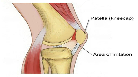
If
injury occurs to the articular cartilage of
the knee, treatment can be more problematic.
There is no widely used, well proven way to
regenerate surface cartilage of significant
damage has occurred. However, treatment with
activity modification, anti-inflammatory
medications, occasionally cortisone
injections, and a variety of developing
arthroscopic surgical techniques is useful
in the majority of patients.

The anterior cruciate ligament is the
main central stabilizer of the knee
during sports. It prevents the shin bone
from sliding forward on the femur bone,
especially when the knee is out
straight. This ligament can be torn
during twisting or other types of
loading maneuvers of the knee. Usually
the athlete feels a pop, and has
significant pain associated with
moderate to severe swelling of the knee.
This can occur in conjunction with other
injuries, such as meniscal tears.
Diagnosis of the tear is made either by
physical examination by the physician,
or by magnetic resonance imaging (MRI)
scan.
If the ligament is completely
disrupted, surgery to reconstruct the
ligament is recommended in athletes who
wish to return to cutting, pivoting, or
jumping sports. This is because you know
that athletes who lack this ligament and
who are engaged in those types of sports
will continue to have "giving out" of
their knee, which can irreparably damage
the cartilage surfaces and lead to early
arthritis. The surgery is performed an
athletes can usually return to play
within 6 to 12 months following injury,
depending upon their recovery
course.

The posterior cruciate ligament is
another central stabilizer of the knee
and prevents the shin bone from sliding
backwards on the femur bone during
sports. It is more important when the
knee is flexed. Injury to this ligament
is much more rare than to the anterior
cruciate ligament.!105BO32.GIF (4620
bytes) In addition, the mechanics of the
ligament are such that unless other
ligaments are injured at the time of PCL
tear, non-operative treatment is usually
pursued. Most patients do very well if
only the posterior cruciate ligament is
torn without having surgery. However, in
patients who have significant symptoms
or other ligament damage, reconstruction
is again performed similar to that for
the anterior cruciate
ligament.

The medial (inner side of the knee) and
lateral (outer side of the knee)
collateral ligaments are additional
ligament structures that prevent the
knee from going into a knock-kneed or
bowlegged posture. Twisting injuries or
being struck on the side of the knee can
partially or completely tear these
ligaments. Low-grade injuries are
treated without surgery. High-grade
injuries, especially if associated with
other ligaments or cartilage damage, may
require surgical treatment.

Kneecap Dislocations are a relatively
common injury in the young athlete. The
rate of dislocation is higher in women
than men. First time dislocations are
usually treated with initial
immobilization followed by
rehabilitation therapy. Recurrent
(multiple) dislocations may require
surgical treatment to repair the torn
tissue that allows the kneecap to keep
dislocating it.

This is a term chopic.GIF (4016
bytes)used to describe wear and tear of
the smooth articular cartilage on the
under surface of the kneecap. This
occurs due to overuse or kneecap
dislocations in athletes. Symptoms
involve pain in the anterior knee with
grinding or catching in the kneecap,
especially with climbing of stairs or
hills, or with jumping and running.
Treatment is often challenging.
Initially, non-operative treatment,
including activity modification, oral
anti-inflammatory medications, and
physical therapy, is undertaken. For
patients in whom this treatment does not
work, occasionally surgery can be of
benefit.

This is a condition of young athletes,
usually male, in which the insertion of
the kneecap tendon into the shin bone
becomes very sore and inflamed. Often a
large bump develops in the bone as well
þ Treatment is usually aimed at
reducing pain and modifying activity
until the symptoms subside. Surgery is
rarely needed.

In General Arthroscopy is the art of
performing surgery in joints with the
use of a small fiberoptic telescope
placed into the joint through small,
quarter- inch incisions. Instruments are
also passed through these incisions to
allow the surgeon to work inside the
knee and perform fine manipulation of
structures in the knee.
The surgeon
(and sometimes the patient) watch the
procedure on a television screen. The
use of arthroscopy has expanded
significantly in the last 10 to 15
years. Currently, we are able to perform
a wide variety of surgical procedures in
both athletes and non-athletes through
the "scope". The advantages of
arthroscopic surgery include the small
incisions, decreased postoperative pain,
and, in some cases, a superior result of
that obtained through a larger "open"
incision.
Some procedures in the
knee that are performed arthroscopically
include:
- Removal of cartilage tears
- Removal of loose bodies in the knee
- Diagnosis of obscure problems of the knee